Tang Dead, Microscope Autopsy. Help ID.

SaltwaterGuruNeeded

Well-Known Member
View Badges
Joined
Jun 29, 2021
Messages
630
Reaction score
275
Location
Tom's River
Rating - 0%
0   0   0
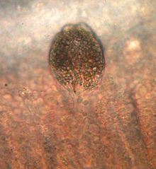
Are there anything in these findings that would've killed my tang? He was breathing kinda fast starting yesterday or the day before...He died this morning. If you have suggestions I could go back and try to get a better picture of something.

@vetteguy53081


IMG_20210826_113733768_HDR~2.jpg
IMG_20210826_114215927~2.jpg
IMG_20210826_115022386~2.jpg
IMG_20210826_115430666~2.jpg


IMG_20210826_121215035~2.jpg

IMG_20210826_122413280_HDR~2.jpg
IMG_20210826_123014246_HDR~3.jpg
IMG_20210826_123426829_HDR~2.jpg
IMG_20210826_123641262_HDR~2.jpg
IMG_20210826_124329697~2.jpg
IMG_20210826_124607097~2.jpg
IMG_20210826_130438124_HDR~2.jpg
IMG_20210826_130922015_HDR~2.jpg
IMG_20210826_131213791~2.jpg
IMG_20210826_132024134~2.jpg


Let me know if I should take more pictures...I still have the fish to take more samples and still have the slides to look back at.
 
OP
OP
SaltwaterGuruNeeded

SaltwaterGuruNeeded

Well-Known Member
View Badges
Joined
Jun 29, 2021
Messages
630
Reaction score
275
Location
Tom's River
Rating - 0%
0   0   0
Pretty awesome of you to do this. Think I maybe see here gill fluke, and a ich cell(maybe). Other maybe pigment cells. To get any real cell damage you might need a little more mag, and cell stains.
Could you point out which is the presumed gill fluke? And suspected ich cell?
 
OP
OP
SaltwaterGuruNeeded

SaltwaterGuruNeeded

Well-Known Member
View Badges
Joined
Jun 29, 2021
Messages
630
Reaction score
275
Location
Tom's River
Rating - 0%
0   0   0
Looking at other thread though. Doesn't seem like a sever amount, could of been a combo of all things combined, and 02. Sorry didn't work out.
Yeah...If other fish(1 goby and 2 clowns) get sick or die, I will fallow my tank. Unless I can eradicate these diseases.

Edit: and yes probably was all them, but to me still a mystery, because I'm not seeing a whole lot of things, that fluke was the only one I saw on the gill sample.

Guessing it's a bacterial infection that killed him, but I still have no clue...
 
OP
OP
SaltwaterGuruNeeded

SaltwaterGuruNeeded

Well-Known Member
View Badges
Joined
Jun 29, 2021
Messages
630
Reaction score
275
Location
Tom's River
Rating - 0%
0   0   0
This is from the Gill squeeze. This is what came out. This is a video.


This one is the tangs Flesh.
 

Jay Hemdal

10K Club member
View Badges
Joined
Jul 31, 2020
Messages
25,950
Reaction score
25,721
Location
Dundee, MI
Rating - 0%
0   0   0
Here is some more...
IMG_20210826_162025862_HDR~2.jpg
IMG_20210826_160937491.jpg
IMG_20210826_160653666.jpg
IMG_20210826_160253064_HDR~2.jpg
IMG_20210826_160110188_HDR~2.jpg
IMG_20210826_155750340~2.jpg
IMG_20210826_155438237.jpg
IMG_20210826_155018037_HDR.jpg
IMG_20210826_153126256_HDR~2.jpg
IMG_20210826_150714590~2.jpg
IMG_20210826_150144595~2.jpg
IMG_20210826_145147223~2.jpg
IMG_20210826_142947701~2.jpg
IMG_20210826_142506079_HDR~2.jpg

Sorry - as I've said, MOTION is the big factor in evaluating disease issues under a microscope. Secondary to that is the magnification factor. You can see all sorts of things at high magnification that are simply artifacts. I remember once, 35 years ago, when I called my curator at the Shedd aquarium over to look at a slide from a dead fish, because I saw a huge amount of round cells and thought it was a disease - he said, well, Jay, what you have there are fish mucus cells.

The only thing I see that gives me pause for thought is the image titled, "tang flesh at 400x". That looks like a type of fluke. Trouble is, the magnification is all wrong - that issue would only look like that at around 80x power.

Microscopes are a great tool at lower power, looking for moving parasites. They fail though, at higher magnifications due to all of the artifacts that show up. The changeover takes place at around 180x. Anything higher than that really needs to be evaluated by tissue sent in for histopathology.

Jay
 
OP
OP
SaltwaterGuruNeeded

SaltwaterGuruNeeded

Well-Known Member
View Badges
Joined
Jun 29, 2021
Messages
630
Reaction score
275
Location
Tom's River
Rating - 0%
0   0   0
Ok, well thanks for the info. I have just attached 2 video's above your post. However videos don't always work on this site. I will try and find a way to use videos with more success in the future. But yes, I understand your point. Nothing really seemed to be alive to me, except at over about 600x and it's possible it's from what you've stated before about the effect of the things moving but they actually arent at higher power.
 

Jay Hemdal

10K Club member
View Badges
Joined
Jul 31, 2020
Messages
25,950
Reaction score
25,721
Location
Dundee, MI
Rating - 0%
0   0   0
This is from the Gill squeeze. This is what came out. This is a video.


This one is the tangs Flesh.

The vibrations of the tiny particles in these videos is the "Brownian motion" I mentioned. I also wonder sometimes if this motion isn't also caused by bacteria (too small to see) bumping into the large particles. In any event, I don't see any pathogens in these videos.

Jay
 

Bubbles, bubbles, and more bubbles: Do you keep bubble-like corals in your reef?

  • I currently have bubble-like corals in my reef.

    Votes: 51 40.8%
  • I don’t currently have bubble-like corals in my reef, but I have in the past.

    Votes: 15 12.0%
  • I don’t currently have bubble-like corals in my reef, but I plan to in the future.

    Votes: 34 27.2%
  • I don’t currently have bubble-like corals in my reef and have no plans to in the future.

    Votes: 23 18.4%
  • Other.

    Votes: 2 1.6%
Back
Top